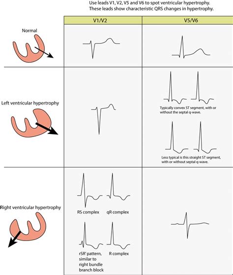
ecg lv hypertrophy | left ventricular hypertrophy life in the fast lane ecg lv hypertrophy Left ventricular hypertrophy changes the structure of the heart and how the heart works. The thickened left ventricle becomes weak and stiff. This prevents the lower left heart chamber from filling properly with blood. Is Louis Vuitton selling pro-Palestinian T-shirts for $800? The internet is divided. People on both sides of the Israeli-Palestinian conflict believe the luxury fashion label may be subtly.
0 · what is lvh on ecg
1 · signs of lvh on ecg
2 · lvh with repolarization abnormality ecg
3 · lv hypertrophy ecg criteria
4 · left ventricular hypertrophy with repolarization abnormality
5 · left ventricular hypertrophy life in the fast lane
6 · ecg showing lvh
7 · ecg in left ventricular hypertrophy
Complete list of doctors in L V Prasad Eye Institute Hyderabad, Hyderabad with their specialities, appointment phone numbers, Email, site, Awards | Sehat
The most common causes of left ventricular hypertrophy are aortic stenosis, aortic regurgitation, hypertension, cardiomyopathy and coarctation of the aorta. . Left ventricular hypertrophy (LVH): Markedly increased LV voltages: huge precordial R and S waves that overlap with the adjacent leads (SV2 + RV6 >> 35 mm). R-wave peak time > 50 ms in V5-6 with associated QRS broadening. LV strain pattern with ST depression and T-wave inversions in I, aVL and V5-6.The most common causes of left ventricular hypertrophy are aortic stenosis, aortic regurgitation, hypertension, cardiomyopathy and coarctation of the aorta. There are several ECG indexes, which generally have high diagnostic specificity but low sensitivity. Left ventricular hypertrophy changes the structure of the heart and how the heart works. The thickened left ventricle becomes weak and stiff. This prevents the lower left heart chamber from filling properly with blood.
Left ventricular hypertrophy (LVH) refers to an increase in the size of myocardial fibers in the main cardiac pumping chamber. Such hypertrophy is usually the response to a chronic pressure or volume load. The two most common pressure overload states are systemic hypertension and aortic stenosis.
Concentric left ventricular hypertrophy is an abnormal increase in left ventricular myocardial mass caused by chronically increased workload on the heart, most commonly resulting from pressure overload-induced by arteriolar vasoconstriction as occurs in, chronic hypertension or aortic stenosis. CONTENTS LAD (left axis deviation) LAHB (left anterior hemiblock) iLBBB (incomplete left bundle branch block) LVH (left ventricular hypertrophy) Diagnostic criteria Interpreting an ECG in the context of LVH LVH versus MI LVH plus ER (early repolarization) Dilated cardiomyopathy HCM (hypertrophic cardiomyopathy) Apical HCM definition of LAD = . Electrocardiogram. Also called an ECG or EKG, this quick and painless test measures the electrical activity of the heart. During an ECG, sensors called electrodes are attached to the chest and sometimes to the arms or legs. Wires connect the sensors to a machine, which displays or prints results.
Current electrocardiographic (ECG) criteria for the diagnosis of left ventricular hypertrophy (LVH) have low sensitivity. Objectives: The goal of this study was to test a new method to improve the diagnostic performance of the electrocardiogram.
Left ventricular hypertrophy can be diagnosed on ECG with good specificity. When the myocardium is hypertrophied, there is a larger mass of myocardium for electrical activation to pass.Left ventricular hypertrophy (LVH) refers to an increase in the size of myocardial fibers in the main cardiac pumping chamber. Such hypertrophy is usually the response to a chronic pressure or volume load. The two most common pressure overload states are . Left ventricular hypertrophy (LVH): Markedly increased LV voltages: huge precordial R and S waves that overlap with the adjacent leads (SV2 + RV6 >> 35 mm). R-wave peak time > 50 ms in V5-6 with associated QRS broadening. LV strain pattern with ST depression and T-wave inversions in I, aVL and V5-6.The most common causes of left ventricular hypertrophy are aortic stenosis, aortic regurgitation, hypertension, cardiomyopathy and coarctation of the aorta. There are several ECG indexes, which generally have high diagnostic specificity but low sensitivity.
Left ventricular hypertrophy changes the structure of the heart and how the heart works. The thickened left ventricle becomes weak and stiff. This prevents the lower left heart chamber from filling properly with blood. Left ventricular hypertrophy (LVH) refers to an increase in the size of myocardial fibers in the main cardiac pumping chamber. Such hypertrophy is usually the response to a chronic pressure or volume load. The two most common pressure overload states are systemic hypertension and aortic stenosis. Concentric left ventricular hypertrophy is an abnormal increase in left ventricular myocardial mass caused by chronically increased workload on the heart, most commonly resulting from pressure overload-induced by arteriolar vasoconstriction as occurs in, chronic hypertension or aortic stenosis. CONTENTS LAD (left axis deviation) LAHB (left anterior hemiblock) iLBBB (incomplete left bundle branch block) LVH (left ventricular hypertrophy) Diagnostic criteria Interpreting an ECG in the context of LVH LVH versus MI LVH plus ER (early repolarization) Dilated cardiomyopathy HCM (hypertrophic cardiomyopathy) Apical HCM definition of LAD = .
Electrocardiogram. Also called an ECG or EKG, this quick and painless test measures the electrical activity of the heart. During an ECG, sensors called electrodes are attached to the chest and sometimes to the arms or legs. Wires connect the sensors to a machine, which displays or prints results.
what is lvh on ecg
signs of lvh on ecg


Current electrocardiographic (ECG) criteria for the diagnosis of left ventricular hypertrophy (LVH) have low sensitivity. Objectives: The goal of this study was to test a new method to improve the diagnostic performance of the electrocardiogram.
Left ventricular hypertrophy can be diagnosed on ECG with good specificity. When the myocardium is hypertrophied, there is a larger mass of myocardium for electrical activation to pass.

lvh with repolarization abnormality ecg
lv hypertrophy ecg criteria
Viesatas upesloku taka. Takas maršruts un sākuma punkts ir mainījušies. Mežs ir ļoti atmosfērisks un Viesatas upe ļoti skaista. Vienā takas posmā pat abās takas pusēs ir pa upei — nepiels posms pirms Viesatā ietek Prūsene.
ecg lv hypertrophy|left ventricular hypertrophy life in the fast lane


